
Guides
Lung nodule grew from 5 mm to 7 mm: real growth or a measurement difference?
A 2 mm change deserves a careful CT comparison. Learn what measurement differences mean and what to prepare for a lung nodule review in China.
By MedicCN, Editorial research · published 2026-10-21 · sources checked 2026-10-21
English article prepared on 21 October 2026 using the linked medical-society, public-health and hospital sources. Source verification is not clinical review: this article has not undergone independent clinical review. Hospital descriptions do not establish a partnership, overseas-patient eligibility or a confirmed appointment.
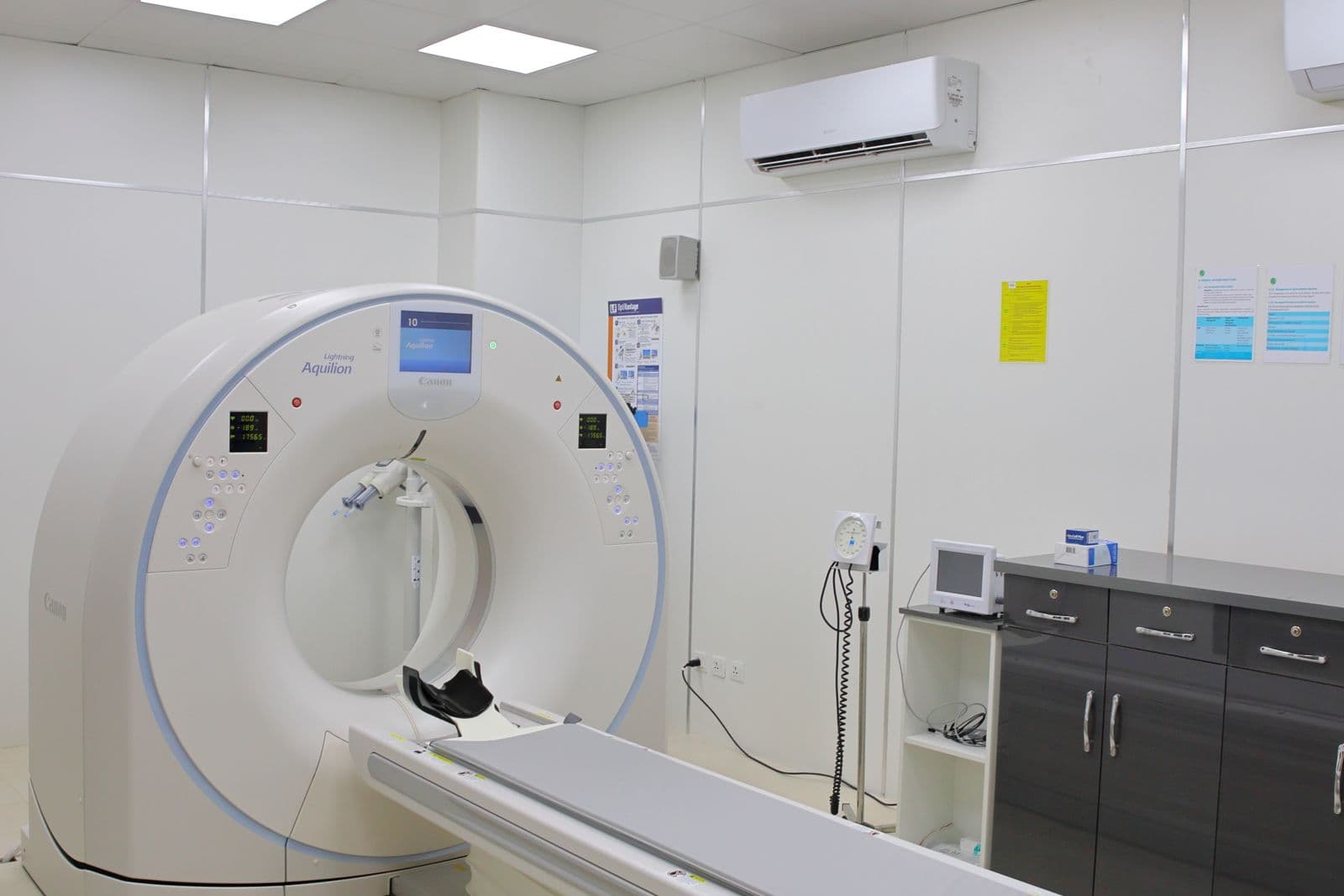
Does 5 mm to 7 mm count as growth?
The latest report says 7 mm; the previous one said 5 mm. That difference needs a radiologist’s review. It may be real growth, but differences in scan technique or measurement can also affect the figures. The original images are needed to tell the difference.
The Fleischner Society’s measurement statement uses a change of at least 2 mm in diameter as a practical threshold for reporting size change. That makes a reported 5-to-7 mm increase worth discussing promptly with the clinician responsible for follow-up. It does not make the two printed numbers a diagnosis or an automatic reason for surgery.
The comparison must concern the same nodule and the same measurement method. Check whether each report gives a longest dimension, an average diameter, a pair of dimensions or the solid component of a part-solid nodule. These are not interchangeable. Ask the radiologist to explain the comparison rather than calculating a risk from the report yourself.
Why can the same nodule measure differently?
Small nodules are sensitive to the way CT images are reconstructed and their borders identified. A different scan technique can make a direct numerical comparison less reliable.
Slice thickness, reconstruction settings and the measurement plane can affect the apparent edge of a nodule. Volumetry research also describes variation between software methods. Thin-section images help the radiologist assess small findings, but thin slices alone do not remove every source of variation.
For illustration, imagine that one report records a longest dimension while another records an average of two dimensions. Even perfectly copied numbers would answer different questions. This is why photographs of selected images are a weak basis for resolving a small reported change: the reviewer needs the full study and its technical information.
| Item | Why it matters |
|---|---|
| Same lesion and location | Multiple nodules can be labelled differently between reports |
| Diameter versus volume | Millimetres and cubic millimetres measure different quantities |
| Whole nodule versus solid component | A part-solid nodule has more than one relevant measurement |
| Scan and reconstruction technique | The visibility of the border affects measurement |
The interval and appearance matter alongside size
The dates of the scans are essential. A change over a short interval and a change across several years need different interpretation; size also needs to be considered with the nodule’s appearance and clinical history.
For some nodules, clinicians use volume measurements and growth-rate estimates to help decide the next step. This is not the same as applying a percentage to a rounded diameter in a report. Changes in a part-solid nodule’s solid component may also matter even when its overall diameter appears similar.
Do not convert a diameter change into a personal cancer probability using an online calculator. The model’s intended population and inputs matter. Ask the clinician whether the evidence supports genuine growth, whether the original follow-up interval should change and what finding would justify an investigation or treatment discussion.
What to send for an image review
Gather both complete CT studies and both reports, plus any older study that already shows the nodule. Keep dates and the original wording intact.
A simple index is enough: examination date, imaging provider, report filename and corresponding image folder. Ask the provider for the original image data, commonly DICOM, rather than exporting a handful of screenshots. Leave the original study structure intact. Include the recommendation made after each scan so the reviewer can see how the plan evolved.
If a report is translated, preserve distinctions such as ‘possible increase’, ‘stable within measurement variation’ and ‘increased solid component’. These phrases should not all become ‘the nodule has grown’. You can flag the sentence you do not understand without replacing it with your own interpretation.
Having the scans reviewed in China
If you are considering a review in China, first ask whether the receiving service can assess the existing images. A repeat scan should follow a clinical decision about what is missing.
Request a pulmonary nodule or thoracic imaging review and state the specific question: ‘Can these studies be compared reliably, and is there convincing interval growth?’ Confirm whether the hospital offers a written imaging opinion, an outpatient assessment or an MDT referral. These are different services and should not be assumed to be available remotely.
Keep your treating clinician informed and retain the existing follow-up appointment unless that team changes it. The useful outcome is a documented interpretation and a next-step plan, including who will arrange and review the next test. Do not let the logistics of a second opinion postpone a time-sensitive local assessment.