
Guides
SMILE vs LASIK vs PRK vs ICL: differences that matter before surgery
SMILE, LASIK, PRK and ICL compared: recovery, tissue changes, prescription ranges, dry eye, night vision and long-term lens risks, with device-specific evidence.
By MedicCN, Editorial research · Medically reviewed by Dr. Jiang Haiyang · published 2026-09-26 · sources checked 2026-09-26
Medically reviewed by Dr. Jiang Haiyang, MedicCN Co-founder and Chief Medical Officer. Sources checked on 26 September 2026; individual suitability and postoperative instructions require assessment by the treating eye-care team.
The main trade-offs at a glance
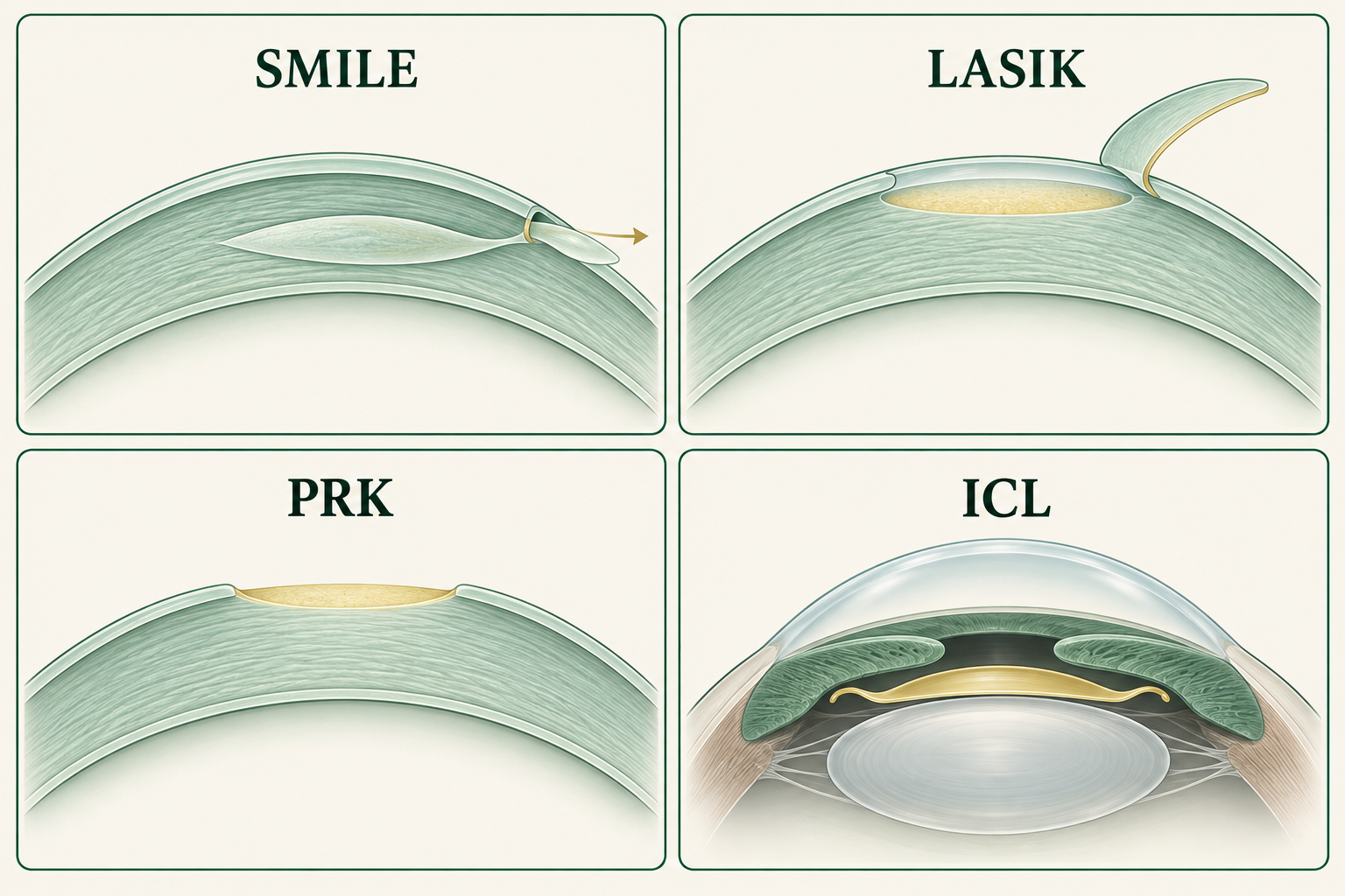
LASIK usually gives the fastest early clarity; SMILE avoids a flap; PRK also avoids a flap but has a more uncomfortable first week. ICL leaves corneal tissue in place and can cover high prescriptions, but adds the risks and monitoring of an implant inside the eye.
| Procedure | How it corrects vision | Early recovery | Distinct trade-off |
|---|---|---|---|
| SMILE | A femtosecond laser creates a corneal lenticule, removed through a small incision. | Useful vision often returns quickly; fine clarity can lag LASIK. | No flap, but tissue removal, dry eye and ectasia risk remain. |
| Femtosecond LASIK | A flap is lifted; an excimer laser reshapes the tissue beneath it. | Often good vision the next day. | Fast recovery, with flap-related trauma precautions and ocular-surface effects. |
| PRK / LASEK / TransPRK | Surface epithelium is removed or moved aside before excimer reshaping. | Usually around a week off work; clarity improves over subsequent weeks. | No stromal flap; more early pain and a risk of corneal haze. |
| EVO ICL / toric ICL | A lens is placed behind the iris, in front of the natural lens. | Early vision can be good, with pressure and position checks still required. | No corneal ablation; cataract, pressure and endothelial-cell risks require ongoing review. |
Prescription ranges are only the first filter
The amount and shape of tissue left after surgery matter more than whether a prescription fits an advertised range. Laser correction changes the cornea’s focusing power; it does not shorten a highly myopic eye or remove retinal disease. ICL supplies optical correction without using corneal tissue, but needs adequate space inside the eye.
LASIK separates a flap before removing stromal tissue. PRK avoids that flap; its epithelial layer regrows, but the reshaped underlying stroma does not. SMILE removes a lenticule under an intact cap except for the access incision. All three therefore require corneal shape and thickness measurements, assessment for keratoconus, and a calculation of the proposed tissue removal. A thin cornea may rule out one plan without making another automatically safe.
The device examples below show why “the LASIK limit” is not one number. Sphere, cylinder and spherical equivalent are different measurements: spherical equivalent is sphere plus half the cylinder. These are US labeling examples, not Chinese approval limits or personal recommendations; local authorization and the exact treatment mode must also match.
EVO’s US requirements include a true anterior-chamber depth of at least 3.0 mm, measured from the back of the cornea to the front of the natural lens, plus an age- and depth-appropriate endothelial-cell count. A measurement taken from the front of the cornea is not interchangeable. A shallow chamber or insufficient cells can exclude ICL even when the cornea is too thin for laser surgery.
| Device / mode | Labeled refractive range | Age |
|---|---|---|
| VisuMax SMILE, 2018 expansion | Sphere −1.00 to −10.00 D; astigmatism treatment cylinder −0.75 to −3.00 D; spherical-equivalent magnitude ≤10 D. | 22+ |
| WaveLight EX500 LASIK, conventional myopia mode | Sphere up to −12 D, astigmatism up to 6 D; guided modes have different limits. | 18+ |
| WaveLight EX500 / Eye-Q PRK | Spherical-equivalent magnitude ≤6 D; sphere ≤6 D and cylinder ≤3 D. | 18+ |
| EVO / EVO+ ICL family | Spherical equivalent −3 to −20 D; correction through −15 D, reduction beyond −15 D. Toric model: cylinder 1–4 D. | 21–60 after FDA supplement S048 |
- FDA — VisuMax SMILE indications, P150040/S003 (2018)
- FDA — WaveLight EX500 LASIK indications, P020050/S006
- FDA — WaveLight PRK indications, P020050/S023 (2016)
- FDA — EVO ICL optical and anatomical indications (2022)
- FDA — EVO ICL age expansion to 21–60, P030016/S048 (2025)
- FDA — corneal and ocular-surface suitability
- FDA — corneal reshaping and refractive errors
Clear distance vision, comfort and night vision recover differently
LASIK’s early speed is a real advantage when rapid visual recovery matters. SMILE’s smaller access incision preserves more early corneal sensation in some comparative studies. Neither feature by itself establishes better final vision or freedom from dry eye.
In a randomized study from Zhongshan Ophthalmic Center, 113 SMILE eyes and 84 femtosecond-LASIK eyes started at approximately −5 D. LASIK gave better day-one uncorrected acuity. At six months, acuity did not differ significantly; SMILE induced less spherical aberration in the 6 mm analysis zone. That optical measurement concerns how peripheral and central rays focus, rather than a universal advantage in every lighting condition.
A separate 40-person trial assigned SMILE to one eye and wavefront-guided LASIK to the other. SMILE eyes retained better corneal sensitivity at months 1, 3 and 6; by month 12 both groups were back to baseline. Patient-reported dry-eye scores did not differ significantly. Existing tear-film or eyelid disease still needs treatment because a disturbed surface affects both comfort and measurement quality.
PRK’s slower start reflects epithelial healing rather than an inherently worse final result. In a 34-person paired-eye randomized study, PRK had worse measured acuity, contrast and visual symptoms at one month; significant differences were no longer found by three months. This older-platform trial explains the recovery trade-off, not the performance of every current laser.
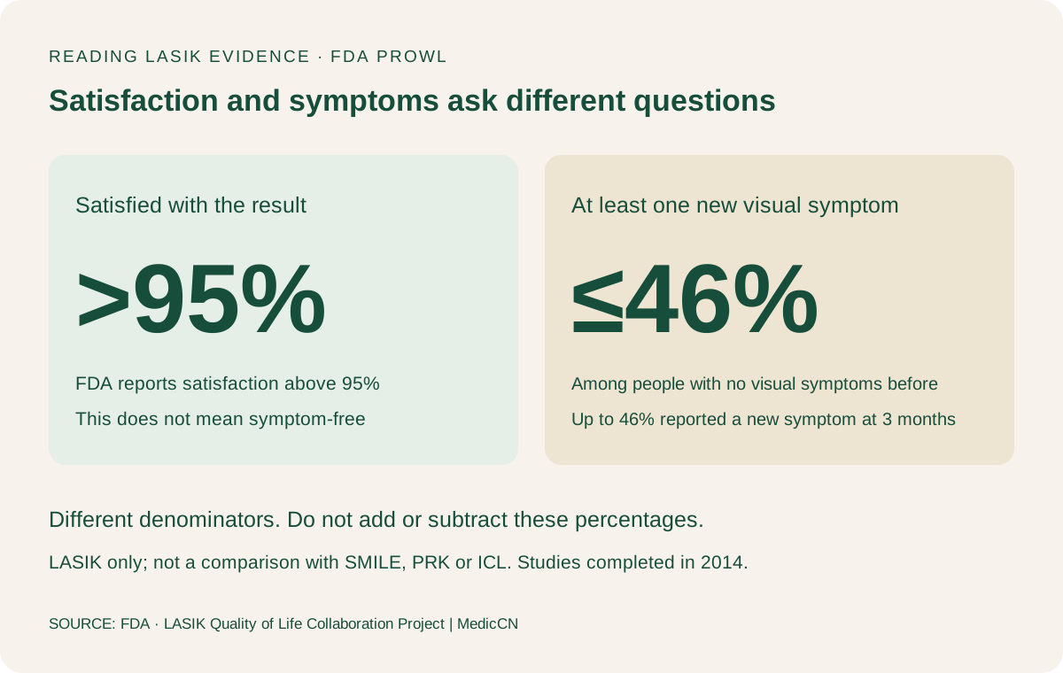
LASIK also has unusually detailed patient-reported data. In FDA PROWL studies, more than 95% were satisfied. Among people without preoperative visual symptoms, up to 46% reported at least one new visual symptom at three months; fewer than 1% reported major difficulty with usual activities because of any one symptom. The chart uses these distinct denominators. A halo can coexist with 20/20 acuity and satisfaction, so night driving requires more than an eye-chart result.
ICL exchanges tissue removal for implant monitoring
ICL is particularly relevant when a large correction would consume too much corneal tissue. Its long-term concerns are different: the lens must remain correctly positioned, fluid must circulate, eye pressure must stay controlled and the corneal endothelium must remain healthy.
The space between the implant and natural lens is called the vault. Excessive vault can narrow the drainage angle; insufficient separation can increase concern about contact with the natural lens and cataract. Other risks include inflammation, infection, glare and a later operation to reposition, exchange or remove the lens. Removal is another operation, not a return to an eye that has never undergone surgery.
In the US prospective EVO study, 327 people aged 21–45 received 629 implants; 579 eyes attended the three-year visit. Mean endothelial-cell density loss was 6.7%. The study reported one anterior subcapsular cataract and two exchanges for high vault. These are eye-level results from a selected, manufacturer-sponsored cohort without a randomized laser-surgery comparator; three years cannot establish lifetime risk.
The FDA has since expanded EVO eligibility to age 60. The 2026 directions for use explicitly state that prospective trial data were not collected in ages 46–60 and highlight greater cataract concern in older and more highly myopic patients. The expanded label should not be confused with new three-year trial evidence in that age group.
Distance correction does not stop the eye ageing
All four approaches can reduce dependence on distance glasses. None restores the youthful focusing ability of the natural lens. Reading glasses can still become necessary as presbyopia develops, and cataract or retinal disease can affect later vision.
A myopic person who currently removes glasses to read may lose that convenience after both eyes are corrected for distance. ICL retains the natural lens, but it does not prevent that lens from stiffening with age. A successful distance result and a new need for readers are therefore compatible outcomes.
Monovision deliberately leaves one eye focused nearer and the other farther. It can reduce reliance on reading glasses, at the cost of binocular depth perception and some low-light performance. A contact-lens trial before permanent correction helps establish whether that compromise is tolerable. For someone who drives at night or needs fine stereoscopic work, distance correction in both eyes with reading glasses may be the more useful target.